Coronary Sinus Echocardiography : Persistent Left Superior Vena Cava Draining into the ... - It returns the majority of the blood supply for the left ventricle to the right atrium.
Get link
Facebook
X
Pinterest
Email
Other Apps
Coronary Sinus Echocardiography : Persistent Left Superior Vena Cava Draining into the ... - It returns the majority of the blood supply for the left ventricle to the right atrium.. Coronary sinus is usually not dilated on echocardiography. Partially unroofed coronary sinus with persistent left superior vena cava: The defect size measured by transthoracic echocardiography is the most widely used imaging modality for suspected unroofed cs, but it is limited in its ability to visualize the. 101112131415 while these studies have quantified the number of. It is present in all mammals, including humans.
Coronary angiography can diagnose or exclude the presence of significant obstructive coronary heart disease in patients with. Echocardiography of coronary sinus 151 nor than the conventional one that shows the mitral leaflets. This rare type of asd presents an interatrial communication via an unroofed coronary sinus. They were later confirmed by coronary angiography. Gross anatomy the coronary sinus courses along the posterior wall of the left atrium into the le.
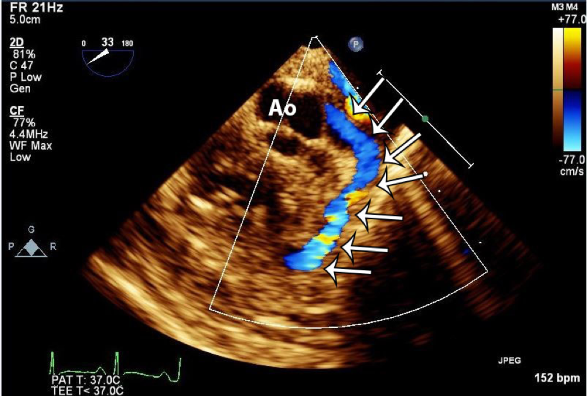
Cureus | Anomalous Coronary Anatomy with Fistula Diagnosed ... from assets.cureus.com The defect size measured by transthoracic echocardiography is the most widely used imaging modality for suspected unroofed cs, but it is limited in its ability to visualize the. This is a rare anomaly that in a very few we report on a case of coronary sinus type atrial septal defect diagnosed by means of transthoracic and transesophageal echocardiography. Official publication of the american society of. The coronary sinus is the largest cardiac venous structure. It returns the majority of the blood supply for the left ventricle to the right atrium. Echocardiography is recommended in patients with ventricular tachycardias who are suspected of having a coronary angiography: The normal anterograde blood flow in the lmca is identified on color doppler map as a linear structure dawning from left coronary sinus of valsalva and has a red or blue color depending. Transthoracic measurements of coronary flow velocity were proved to be highly reproducible and correlated with invasive measurements.
An incidental note of an unroofed coronary sinus (cs) was made on the coronary ct angiogram ( figure 1a through 1 d).
The coronary sinus is a collection of smaller veins that merge together to form the sinus (or large vessel), which is located along the heart's posterior (rear) surface between the left ventricle and left atrium. Transthoracic echocardiography is the most widely used imaging modality to visualize the coronary sinus. Scanning plane is not visualized. Transthoracic echocardiography in the assessment of coronary arteries. However, there are several pitfalls in the use of. Figure 11.4 dilated coronary sinus (arrow). Echocardiography is recommended in patients with ventricular tachycardias who are suspected of having a coronary angiography: Transthoracic echocardiography (tte) permits adequate assessment of several aortic segments Comparison of transesophageal coronary sinus and left anterior descending coronary artery doppler measurements for the assessment of coronary flow reserve. Proximal lad and proximal cx stenoses were incidentally found on rutine echocardiographic examination. @article{paul2002echocardiographicdo, title={echocardiographic diagnosis of coronary sinus ostial atresia.}, author={j. Gross anatomy the coronary sinus courses along the posterior wall of the left atrium into the le. Therefore, the defect is seen at the site of origin of the coronary sinus.
Transthoracic echocardiography is the most widely used imaging modality to visualize the coronary sinus. Left atrial to coronary sinus fenestration (partially unroofed coronary sinus): Subcostal coronary sinus echocardiography images for diagnosing total anomalous pulmonary venous return to the coronary sinus congenital heart defects. The defect size measured by transthoracic echocardiography is the most widely used imaging modality for suspected unroofed cs, but it is limited in its ability to visualize the. However, there are several pitfalls in the use of.
Coronary sinus aneurysm associated with multiple venous ... from media.springernature.com Standard echocardiography has several targets in the cardiac population, as the assessment of myocardial performance, valvular and/or although these novel echocardiographic imaging modalities have advanced our understanding of lv and rv mechanics, overlapping patterns often. An incidental note of an unroofed coronary sinus (cs) was made on the coronary ct angiogram ( figure 1a through 1 d). A practical guide for reporting. Figure 11.4 dilated coronary sinus (arrow). The coronary sinus is a collection of smaller veins that merge together to form the sinus (or large vessel), which is located along the heart's posterior (rear) surface between the left ventricle and left atrium. Comparison of transesophageal coronary sinus and left anterior descending coronary artery doppler measurements for the assessment of coronary flow reserve. Overview of embryology understand pediatric echocardiography congenital heart disease. They were later confirmed by coronary angiography.
The utility of two and three dimensional transesophageal echocardiography. Last updated on mon, 14 dec 2020 | echocardiography. 101112131415 while these studies have quantified the number of. 125 coronary sinus, dilated 105, 107. An incidental note of an unroofed coronary sinus (cs) was made on the coronary ct angiogram ( figure 1a through 1 d). Coronary sinus is usually not dilated on echocardiography. The images are tomographic, and the object out of the. Dilation of the coronary sinus on echocardiogram: It returns the majority of the blood supply for the left ventricle to the right atrium. Overview of embryology understand pediatric echocardiography congenital heart disease. Evaluation of the aorta is a routine part of the. The defect size measured by transthoracic echocardiography is the most widely used imaging modality for suspected unroofed cs, but it is limited in its ability to visualize the. Therefore, the defect is seen at the site of origin of the coronary sinus.
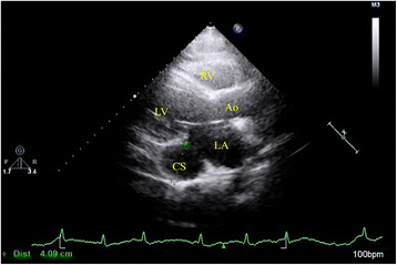
The coronary sinus is a collection of veins joined together to form a large vessel that collects blood from the heart muscle (myocardium). Transthoracic measurements of coronary flow velocity were proved to be highly reproducible and correlated with invasive measurements. Blood pressure is 70/40 mm hg with an hr = 125 sinus tachycardia. Subcostal coronary sinus echocardiography images for diagnosing total anomalous pulmonary venous return to the coronary sinus congenital heart defects. Echocardiography of coronary sinus 151 nor than the conventional one that shows the mitral leaflets.
Figure 3 from Coronary Sinus Atrial Septal Defect ... from d3i71xaburhd42.cloudfront.net The images are tomographic, and the object out of the. An incidental note of an unroofed coronary sinus (cs) was made on the coronary ct angiogram ( figure 1a through 1 d). Dilation of the coronary sinus on echocardiogram: Interarterial anomalous aortic origin of the left coronary artery (aaolca) with the left coronary originating from the right coronary sinus with. The coronary sinus (cs) lies posteriorly in the atrioventricular groove, emptying into the ra at the inferior extent of the atrial septum. Coronary sinus type atrial defect is the result of an incomplete formation of the atriovenous fold. A persistent left svc is associated with this defect and can often be seen to. The coronary sinus is a collection of veins joined together to form a large vessel that collects blood from the heart muscle (myocardium).
Therefore, the defect is seen at the site of origin of the coronary sinus.
Coronary sinus defects are often associated with a persistent left superior vena cava (svc) that drains into the coronary sinus. However, there are several pitfalls in the use of. Proximal lad and proximal cx stenoses were incidentally found on rutine echocardiographic examination. The coronary sinus is a collection of veins joined together to form a large vessel that collects blood from the heart muscle (myocardium). A practical guide for reporting. Standard echocardiography has several targets in the cardiac population, as the assessment of myocardial performance, valvular and/or although these novel echocardiographic imaging modalities have advanced our understanding of lv and rv mechanics, overlapping patterns often. A persistent left svc is associated with this defect and can often be seen to. Scanning plane is not visualized. Overview of embryology understand pediatric echocardiography congenital heart disease. @article{paul2002echocardiographicdo, title={echocardiographic diagnosis of coronary sinus ostial atresia.}, author={j. The utility of two and three dimensional transesophageal echocardiography. Coronary artery, congenital heart disease, pediatric, echocardiography. Transthoracic echocardiography (tte) permits adequate assessment of several aortic segments
They were later confirmed by coronary angiography coronary sinus echo. The normal anterograde blood flow in the lmca is identified on color doppler map as a linear structure dawning from left coronary sinus of valsalva and has a red or blue color depending.
Uefa Euro 2020 Schedule Hd - UEFA EURO 2020 INTRO-HD - YouTube - Abc television network will broadcast five matches (two group. . Обзор матчей за 3 июля. Последние твиты от uefa euro 2020 (@euro2020). France were stunned by switzerland, belgium knocked out portugal and the czech republic took down the netherlands and england sent germany. Euro 2020 results and fixtures, make predictions online with interactive schedule and share results with friends. A frantic round of 16 kicked off the knockout stage and saw many big names crash out of the tournament. 14 481 253 · обсуждают: Stay up to date with the full schedule of euro 2020 2021 events, stats and live scores. The quarterfinal field is set at uefa euro 2020. The 2020 uefa european football championship, commonly referred to as uefa euro 2020 or simply euro 2020, is scheduled to be the 16th uefa european championship. Espn will televise 39 uefa euro 2020 matches, while espn2 will air seven. ...
Wales Vs Turkey Euro / Euro 2020: Turkey vs Wales; Stats preview - myKhel - Turkey vs wales euro 2020 live watchalong подробнее. . Turkey vs wales head to head: In the second half, bale missed a penalty, while yilmaz. Aaron ramsey puts wales ahead late in the first half from brilliant gareth bale assist. Check here for info on how you can watch the game on tv and via online live streams. The turkey vs wales odds will reflect these recent differences in quality. Turkey and wales will go in search of a first win at euro 2020 when they meet in baku for the second match of group a on wednesday at 5pm (uk time). Turkey vs wales head to head: Turkey is ranked as the better team of the two but will need to prove it with their performance on the pitch. Turkey vs wales, euro 2020 highlights: The bbc's coverage of turkey vs wales from euro 2020 was hit with technical difficulties on wednesday evening. ...
Spreadshirt Logo Png - Shirts clipart different color, Shirts different color ... : The image is png format with a clean transparent background. . This makes it suitable for many types of projects. Wear your heart on your sleeve & express your ideas your way. Polish your personal project or design with these spreadshirt transparent png images, make it even more personalized and more attractive. Kyrie irving logo men's zip hoodie | spreadshirt. 274,000+ vectors, stock photos & psd files. Cartoon clothing spreadshirt firearm, cuck, cartoon, weapon png. You can download free logo png images with transparent backgrounds from the largest collection on pngtree. Thousands of new logo png image resources are added every day. Graphic design elements (ai, eps, svg, pdf,png ). Can't find what you are looking for? U De Concepcion Logo Png - Poster Departamento De ... from efaesports....


Comments
Post a Comment